Measurement of Emission Spectrum of Fluorescence Microbeads using uSight-2000
The demand for miniaturization has mandated that the dimension of colour pixels and photonic devices to be scaled down corresponding. For example, the latest smartphone’s CCD sensor size has decreased to as small as 1 micron. Therefore, it becomes more challenging to accurately determine the photon interaction of such small-scale photonic device at high spatial resolution, including colour, absorption and/or emission properties. [1,2]
uSight-2000 is designed to fulfil such demand with affordable cost. uSight-2000 micro-spectroscopy system that provides very good spatial resolution and ensures accurate spectrum collection from a very small area of interests. With its unique LED position indicator approach, users can easily verify the exact collection spot position and dimension with accuracy down to diffraction limit of light.
In this application note, we demonstrate uSight-2000 capability to measure fluorescence spectra from microbeads loaded with multiple dyes with excellent spatial resolution.
The entire integrated system consists of a uSight-2000 module mounted onto a Nikon Ci-S microscope equipped with a motorized stage, fluorescence light source from 89 North and our standard integrated USB3.0 camera to capture the fluorescence image. We used 1.0 µm TetraSpeck™ microspheres and a 400 nm long pass filter to remove the UV excitation light. All measurement were taken with Nikon 100X 0.9 NA objective. The uSight-2000 offers UV-Vis-NIR measurement which is sensitive enough for fluorescence spectrum measurement with excellent spatial resolution.
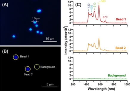
Figure 1. (A-B) Fluorescence image and (C) its respective spectrum of TetraSpeckTM microbeads
Figure 1(A) is an example of fluorescence image taken from TetraSpeckTM microbeads. The average size of the microbead is around 1.5 ± 0.05 µm (statistic collected from >10 microbeads). These TetraSpeckTM microbeads are loaded with 4 different dyes with well-separated excitation/emission peaks, i.e. 360/430 nm (blue), 505/515 nm (green), 560/580 nm (yellow) and 660/680 nm (dark red), respectively.
We collected the fluorescence spectra from 3 different locations from the sample, as shown in Figure 1(B). Figure 1 (C) shows the spectrum from the microbeads (bead 1 and 2) contains multiple emission peaks which correspond to 436, 476, 510, 580 and 670 nm. One important advantage is the good spatial (x, y) resolution from the uSight-2000. The emission spectrum collected from Bead 2 and background in Figure 1C are less than 5 µm apart with no spectral cross-talk is observed. In fact, we measured with NIST traceable reference standard all our uSight-2000 systems to show the achievable spatial resolution is less than or equal to 1µm[3] when using 100x objective lens.
In conclusion, uSight-2000 is UV-vis-NIR microspectroscopy that enables various spectroscopic measurements with high spatial selectivity ranging from colour, reflectance, absorption, transmission, emission (fluorescence) and scattering measurements. uSight-2000 is an affordable spectroscopy solution that can even be incorporated to your existing microscope to further strengthen your research capability.
Reference:
[1] Nano Letters, 2014, 14, 4023-4029. [2] Nature communications, 2014, 5: 5361.
[3] Spatial resolution of each Microspectroscopy system will vary around 15-20% of the designed specification due to compounded mechanical tolerance of lens and other components by the respective microscope manufacturer. Nevertheless, all our complete uSight-2000 system that comes complete with a microscope will be pre-calibrated with a NIST traceable standard to reflect the exact dimension of our measurement spot against the available objective lens. Typically using a 100X lens on a Nikon microscope will yield a 1mm (+/-0.1 mm) measurement spot, whereas on an Olympus microscope with equivalent parts the spot will be 0.9um (+/-0.1 mm).
