Raman mapping for single layer graphene
The production of large scale and defect-free single-layer graphene is in high demand for their emerging applications in solar panel, light emitting devices, and etc.. However, it remains technically challenging to produce defect-free graphene at large scale. Very often, domains of defects and/or multi-layered graphene are present, and significantly compromise the performance of graphene.
Hence, quality assessment of commercial graphene is a major concern for many end users. Raman spectroscopy, a high throughput and non-destructive spectroscopic technique, can provide a quick and simple identification of the quality of the graphene. The quality of graphene can be assessed by the intensity ratio of G’/G bands (2680 cm-1/1580 cm-1), which serves as an indicator of the presence of single or multiple-layer graphene. In this application note, we demonstrate the combination of Raman mapping and statistical analysis as a powerful method to better assess the quality of graphene and the distribution of defects or multi-layer on a sample.

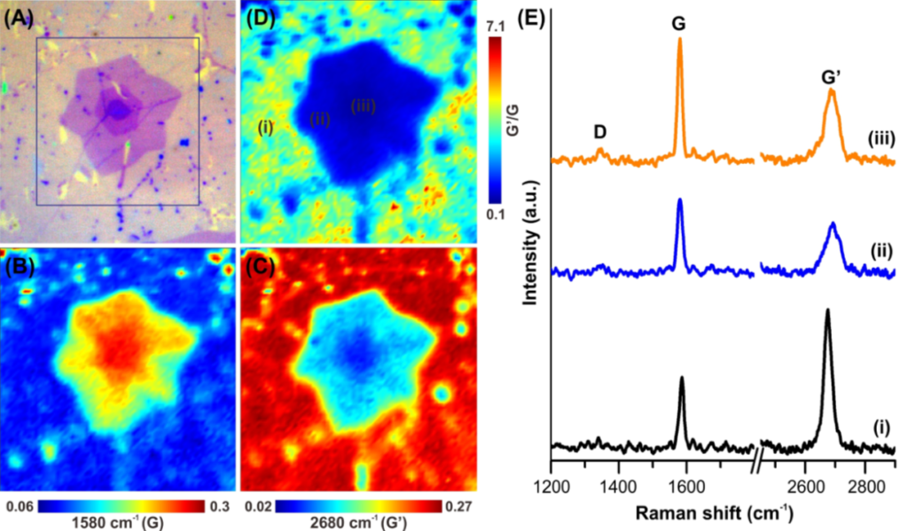
Figure 1 Raman mapping of a transferred graphene sample. (A) optical image, Raman map at (B) 1580 cm-1 (G-band), (C) 2680 cm-1 (G’-band), and (D) intensity ratio of 2680 cm-1/1580 cm-1. (E) Raman spectra from location (i-iii) at (D).
Raman mapping was performed using uRaman-532 on Nikon Ci microscope from Technospex Pte. Ltd. The objective used was 100X with N.A. 0.9 (Nikon Plan Fluor) and motorized stage was used for sample scanning. The graphene sample is fabricated using a CVD growth method on copper film, which consists mostly of a single layer graphene with defects. The graphene is subsequently transferred to 280 nm SiO2 on silicon substrate. [1]
We performed a Raman mapping on a hexagonal shaped region on the graphene sample, as highlighted in a blue box in Figure 1(A). The Raman intensity maps of G (1580 cm-1) and G’ (2680 cm-1) bands show inhomogeneous distribution of Raman intensities in Figure 1(B) and (C), respectively. This suggests the presence of defects. Indeed, we observe three different types of Raman spectra collected from three different locations of the scanned region (i-iii in Figure 1(D) and (E)). The results indicate the presence of various layers of graphene in the sample.
Next, we performed hierarchical cluster analysis (HCA) to determine the hierarchy and distribution of Raman spectral clusters on the Raman map. HCA is a statistical method that groups a set of similar spectra into same group (known as “cluster”). Hierarchical cluster analysis is one of the methods that differentiate spectra based on “distance”, i.e. similarity of each spectrum from its closer to further. The algorithm connects the “spectra” to form a “cluster” based on their “distance”. [2] We used CytoSpec software to perform Hierarchical Cluster Analysis (HCA) of the Raman map we obtained in Figure 1.

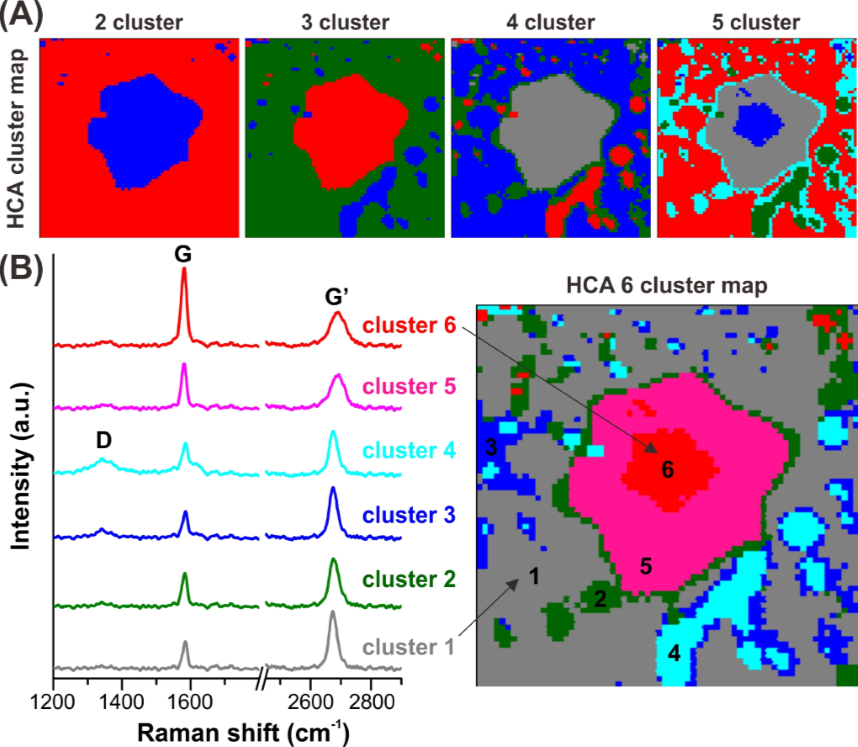
Figure 2 (A) HCA cluster map and (B)average spectra from different cluster corresponding to its HCA cluster map.
HCA cluster maps clearly show different types of clustering from the Raman image. When the number of cluster is increased from 2 to 6, more detailed individual clustering can be observed on the map (Figure 2). For example, the 6-clustered map (Figure 2B) illustrates the distribution of different clusters and their corresponding average spectra. The HCA analysis indicates that among the 6 clusters identified, cluster 1 – 4 can be grouped as single layer of graphene. Their clustering is due to the different degree of defects presence. The defect layers become more significant from cluster 1 – 4, as illustrated by the increasing prominence of D-band (cluster 4). On the other hand, cluster 5 and 6 can be correlated to multi-layers of graphene, i.e. 2L and 3L of graphene, respectively. This is based on the increasing strong G band’s intensity as the number of graphene layer increases (Figure 2B -spectra). In overall, the HCA can segmentize the composition of the graphene sample by analyzing the differences of the hyperspectral images of the sample.
HCA is powerful method to breakdown a complicated Raman map and organize it into clusters of similar feature. In this example, we show only 6 clustering of a graphene sample, but users can use higher number of clusters if the sample is highly inhomogeneous. HCA map is complementary to intensity mapping, but the cluster image provides better visualization of defects distribution and areas with multi-layers graphene. This provides invaluable information for users to quickly assess the quality of graphene layer on their sample.
Acknowledgement
We thank Dr. Peter Lasch, the creator of CytoSpec in providing evaluation copy of CytoSpec 2.00.04 for HCA analysis performed in this application note. We thank Mr. Calvin Wong (NUS) for providing the graphene sample for measurement.
Reference
[1] ACS AMI 2014,6,20464; [2] Nature Protocol 2014,9,1781
